2026 · Published
Research
Cervical Spine Imaging
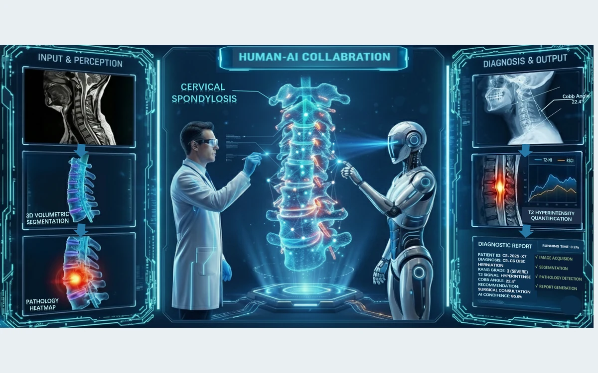
Interpretable cervical MRI analysis spanning anatomical segmentation, clinical measurements, and spinal-cord T2 hyperintensity detection.
Clinical motivation
Cervical spondylosis assessment requires consistent interpretation of multiple anatomical structures, compression patterns, and signal abnormalities.
Technical challenges
- Cervical MRI combines multiple small anatomical structures with heterogeneous degenerative changes.
- Signal abnormalities may be sparse, while scanner and acquisition differences alter normal appearance.
- Routine anisotropic scans limit reformatted views and three-dimensional quantitative assessment.
Research progression
- Pathology-guided segmentation and automated cervical imaging indicators
- Uncertainty-aware detection of spinal-cord T2 hyperintensity
- Isotropic 3D cervical MRI reconstruction from routine scans
- Systematic synthesis of artificial-intelligence tasks and modalities in degenerative cervical myelopathy imaging
Current directions
- Multimodal cervical imaging and structured quantitative analysis
- External validation across acquisition settings
- Uncertainty-aware review of automated measurements
Related publications
Representative publications
2026 · Published
Pathology-Guided AI System for Accurate Segmentation and Diagnosis of Cervical Spondylosis
2026 · Published
U2AD: Uncertainty-based unsupervised anomaly detection framework for detecting T2 hyperintensity in MRI spinal cord
2026 · Under Review